


Interleukin-2 (IL-2): A Multifunctional Biomolecule
IL-2 and IL-2 Receptor Series: Pioneering Solutions for Immunotherapy Research
Interleukin-2 (IL-2) is a 15kDa secretory protein with a 4-alpha-helical bundle structure. As a T-cell related growth factor, IL-2 can enhance the killing activity of NK cells and promote B cells to produce immunoglobulins. In addition, it aids in the development of regulatory T cells (Tregs), thereby producing peripheral T cell immune tolerance, as well as regulating the proliferation and differentiation of activated T cells. In summary, IL-2 has multiple biological functions.
The IL-2 Receptor Family
IL-2, as an important immunoregulatory cytokine, is primarily produced by activated CD4+ T cells and can also be stimulated to be produced by innate lymphoid cells, activated CD8+ T cells, dendritic cells, B cells, and mast cells. In this process, the secretion of IL-2 is regulated by several key transcription factors (including AP-1, NFκB, NFAT), and it functions through dimeric or trimeric IL-2 receptors (IL-2R) [1].
IL-2R is a heterotrimer composed of alpha, beta, and gamma chains. All receptors signal through JAK1 and JAK3, activating downstream STATs factors such as STAT5. The three reported types of IL-2R subunit deficiencies are all related to immunodeficiency and immune dysregulation/autoimmune diseases. IL-2R-related diseases first came into public view with a patient having an IL-2R gamma (CD132) deficiency, clinically manifested as a severe T-B+NK- combined immunodeficiency (X-SCID). Later, cases of human IL-2R alpha (CD25) and IL-2R beta (CD122) deficiencies were discovered [2]. IL-2R gamma is not only a receptor for IL-2 but also for IL-4, IL-7, IL-15, and IL-21. IL-4 has the function of activating B cell proliferation and inducing CD4+ T cells to differentiate into Th2 cells; IL-7 has varying degrees of impact on T cell survival and proliferation; IL-15 provides early stimulation for T cell proliferation and activation, and it can block IL-2 induced T cell apoptosis, which is crucial for maintaining long-term immunity levels [3].
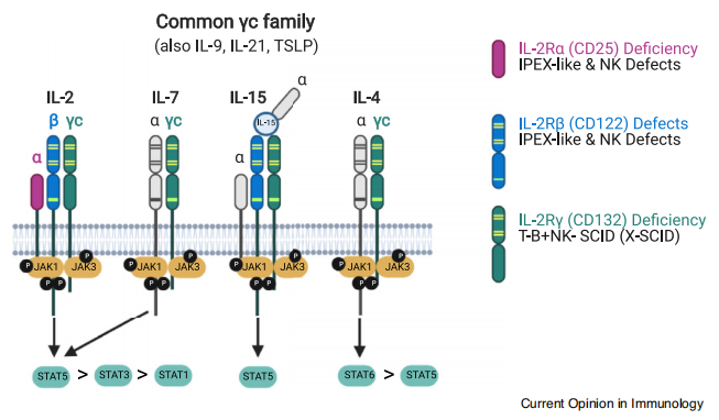
Figure 1. IL-2 and its receptor family [2]
IL-2 signals through heterodimerization of the cytoplasmic domains of IL-2R beta and gamma c, mainly activating three signaling pathways: phosphatidylinositol 3-kinase (PI3K)/AKT, Ras-MAP kinase, and the JAK-STAT pathway (Figure 1). These three signaling pathways collectively mediate cell growth, survival, activation, and the induction of differentiation and death.
Although each IL-2R subunit can be expressed on T cells and NK cells, the effect of each subunit on downstream signal transduction and cell function depends on the level of transcriptional regulation and the affinity for IL-2. IL-2 has multiple targets, including CD4+ T cells, CD8+ T cells, B cells, and NK cells, and sometimes plays opposing functions in adaptive immune responses: on one hand, IL-2 promotes the differentiation of CD4+ T cells into Th1 and Th2 cells, but inhibits the differentiation of CD4+ T cells into inflammatory Th17 cells. IL-2 promotes the activation and formation of memory phenotypes in CD4+ and CD8+ T cells. On the other hand, IL-2 also promotes the apoptosis of activated T cells, which in turn limits the proliferation of effector T cells, constituting negative feedback regulation. Deficiencies in the IL-2R alpha and IL-2R gamma subunits disrupt this IL-2-mediated feedback regulation, ultimately leading to the occurrence of autoimmune/inflammatory diseases.
IL-2 regulates innate immune cells
In humans, monocytes express IL-2R under the stimulation of IFN-γ, but only activated monocytes can bind with IL-2. During the inflammatory response, the expression of IL-2R will affect macrophage differentiation, limiting T cell proliferation and function. The use of IL-2 alone, or in combination with other cytokines such as TNF-α or IFN-γ, can increase the cytotoxicity of macrophages. Dendritic cells can express all three subunits of IL-2R. They translocate IL-2R alpha (CD25) to the initial T cells that need to bind with high affinity to IL-2, causing them to differentiate into cells highly expressing IL-2R alpha (CD25) CD4+ cells and CD8+ T cells (Figure 2).
Most IL-2 receptors on the surface of NK cells are dimers with low affinity, so NK cells often compete with other cells (such as dendritic cells) for IL-2. This competitive mechanism leads to the possible inhibition of NK cells by mesenchymal stem cells, Treg cells, or CD8+ T cells. Innate lymphoid cells (ILCs) are distributed in all organs, regulating the immune response to environmental stimuli. Based on surface molecules, the ability to produce different cytokines, and signature transcription factors, ILCs have been divided into three main subgroups: T-bet+ ILC1s that produce IFN-γ, GATA3+ ILC2 that produce IL-5 and IL-13, and RORγt+ ILC3 that produce IL-17 and IL-22. IL-2 effectively promotes the production and proliferation of ILC2 both in vivo and in vitro [1].
IL-2 regulates adaptive immune cells
CD4+ T cells act as helper cells, executing a series of functions by differentiating into specific subgroups after exposure to antigen stimulation and responding to cytokine signals. These functional subgroups mainly include TH1, TH2, TH17, TFH (Follicular Helper T cells), and Treg (Figure 2). During the differentiation of TH1 cells, IL-2 induces the production of IFN-γ and enhances the expression of IL-12 Rβ2 and T-bet. This regulation depends on the direct binding of IL-2 induced STAT5a/STAT5b with IL12rb2/Tbx21 sites. In addition, IL-2 enhances the expression of MYC and HIF1α, thereby promoting glycolysis in CD4+T cells [1]. IL-2 can maintain the pluripotency of CD8+ T cells by promoting the expression of TCF1, demonstrating powerful anti-tumor functions; however, with the progression of the tumor, IL-2 also mediates the exhaustion of CD8+ T cells [3]. IL-2 is an essential factor for Treg regulatory cell differentiation, immune level, and maintenance of homeostasis. Treg cells control the inflammatory response, their efficiency decreases in inflammatory tissues, and they may even become unstable due to the loss of FOXP3 expression, and transform into a phenotype with more traditional CD4+T cell characteristics, referred to as "ex-T cells" [4].
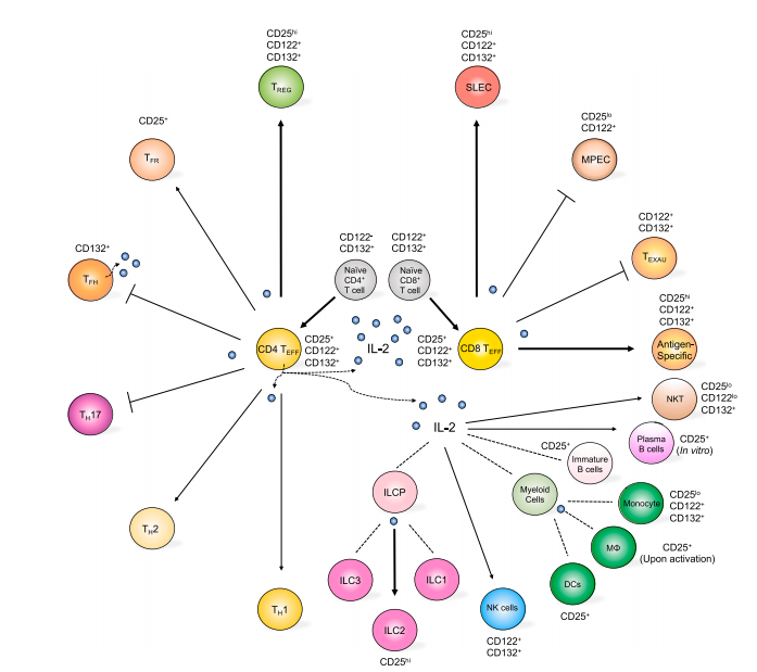
Figure 2. IL-2's role in stimulating the differentiation of immune cells [1] (The arrows represent the positive regulation of specific cell type differentiation mediated by IL-2; bold arrows indicate strong promotion; dashed lines represent functions of IL-2 that are not yet clear)
Due to the important role of IL-2 and IL-2R in maintaining immune tolerance and host immunity, more and more new research is focusing on novel treatment plans for IL-2 related diseases, which is conducive to the rapid expansion of the field of immunotherapy. Early clinical trial results suggest that low-dose IL-2 treatment has a certain safety and efficacy in terms of disease attacks in autoimmune patients [5].
Beta LifeScience provides IL-2 and its receptor proteins of different species and tags, which helps the development of IL-2 drugs and cell therapy drugs.
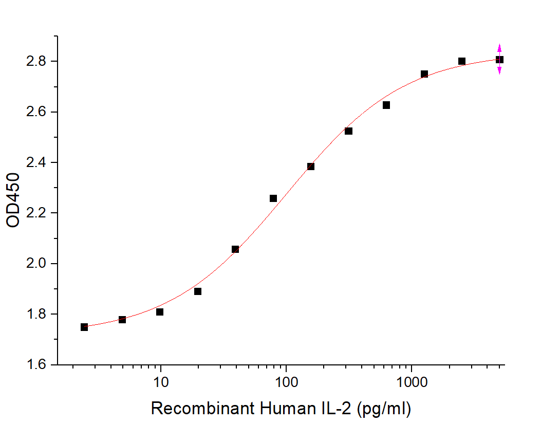
Featured Product 1: Recombinant Human IL-2
Product Features:
- Designed for cellular therapy applications
- Expressed in E. coli, tag-free
- Produced using non-animal source materials and pharmaceutical excipients
- Sterile, free of aminobenzyl penicillin, no HBV, HCV, HIV and other pathogens, mycoplasma-free
- Endotoxins less than 10EU/mg, host protein residues less than 0.005%, exogenous DNA residues less than 10ng/mg
- High purity, good activity, high stability
- Good batch consistency
- Pharmaceutical vial packaging
Applications:
- T cell growth factor, promotes T cell survival, enhances the killing activity of T cells
- Can promote the proliferation of NK cells and maintain long-term growth of NK cells
- Can promote the survival, expansion, and activation of LAK/TIL cells in vitro
- Can promote B cells to express IL-2R, promoting B cell proliferation and immunoglobulin production
- Activates the cytotoxicity of monocytes and macrophages
- Increases the levels of other factors in serum, including IFN-γ, TNF-α/β, and other cytokines, used to regulate immune response
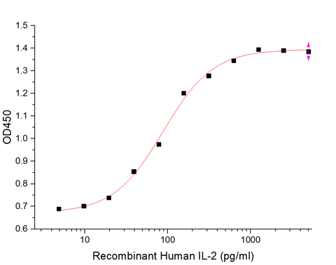
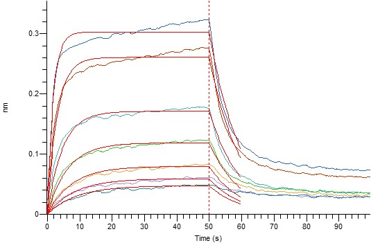
Data Display:
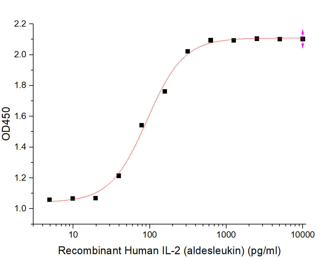
Featured Products 2: IL-2 Series
Product Features:
- Various species: Human, Mouse, Rat, meeting the needs of different species cell culture
- T cell activity proliferation verification, high activity
- Multiple tags available: Tag-free, His tag, and Avi tag
- Various packaging sizes to choose from (10ug/50ug/500ug/1mg)
Applications:
- T cell growth factor, promotes T cell survival, enhances the killing activity of T cells
- Can promote the proliferation of NK cells and maintain long-term growth of NK cells
- Can promote the survival, expansion, and activation of LAK/TIL cells in vitro
- Can promote B cells to express IL-2R, promoting B cell proliferation and immunoglobulin production
- Activates the cytotoxicity of monocytes and macrophages
- Increases the levels of other factors in serum, including IFN-γ, TNF-α/β, and other cytokines, used to regulate immune response
- IL-2 receptor binding verification
- IL-2 Superkine enhances the affinity to IL-2RB, eliminates the dependence of IL-2 on IL-2RA expression, induces stronger cytotoxic T cell proliferation, improves in vivo antitumor response
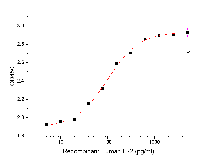
Data Display:
|
Cat. No. |
Product Name |
Expression System |
|
Recombinant Human IL-2(Tag-free) |
E.coli |
|
|
BL-1777NP |
Recombinant Human IL-2 aldesleukin |
E.coli |
| BL-2899NP |
Recombinant Human IL-2 Superkine (C-6His) |
Human Cells |
|
Recombinant Human IL-2 (C-6His) |
Human Cells |
|
| BL-2730NP |
Biotinylated Human IL-2 (C-6His-Avi) |
Human Cells |
|
Recombinant Mouse IL-2(Tag-free) |
E.coli |
|
|
Recombinant Rat IL-2 (C-6His) |
Human Cells |
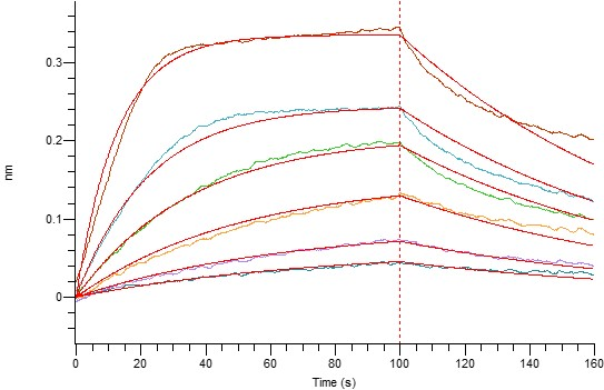
Featured Products 3: IL-2 Receptor Series
Product Features:
- All are mammalian cell-expressed, high purity
- Affinity verification, IL-2 with IL-RA, IL-2RB and IL-2RG affinity weakens in sequence, consistent with IL-2 binding rules with different receptor subtypes
- Different species and receptor subtypes available to choose from
- Various tags including His, Fc, and Avi tag
- Various packaging sizes to choose from (10ug/50ug/500ug/1mg)
Applications:
- Research of IL-2 affinity with different receptor subtypes
- Animal immune preparation of antibodies
- In vitro antibody screening and function validation
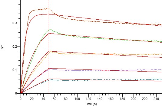
Data Display:
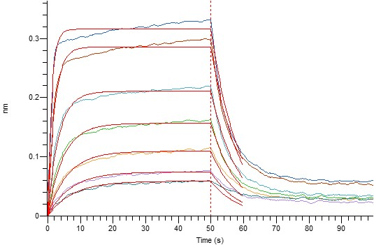
BLI (BL-0100NP: IL-2RA-Fc and IL-2 Affinity Data)
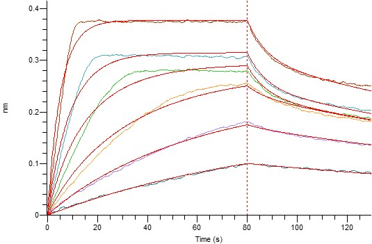
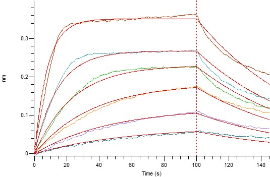
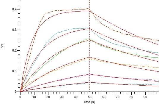
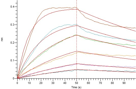
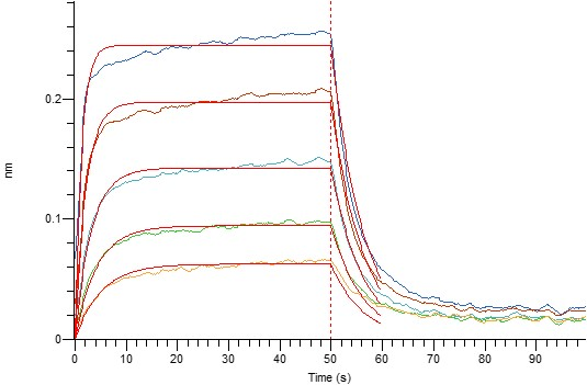
BLI (BL-0102NP:IL-2RB-Fc and IL-2 Affinity Data)
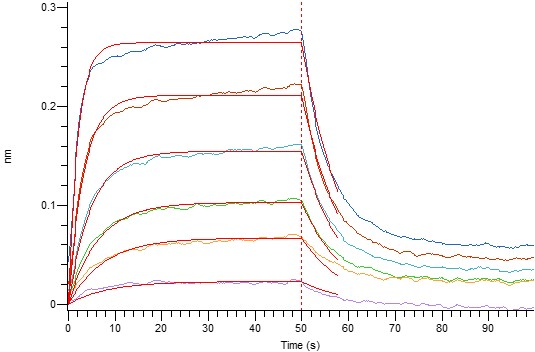
Loaded Human IL-2RB-Fc(Cat#BL-0102NP) on Protein A Biosensor, can bind Human IL-2(Cat#BL-1714NP) with an affinity constant of 0.29 uM as determined in BLI assay.
|
Cat. No. |
Product Name |
Expression System |
|
Recombinant Human IL-2RA/CD25 (C-6His) |
Human Cells |
|
|
Recombinant Human IL-2RA/CD25 (C-Fc) |
Human Cells |
|
|
Recombinant Mouse IL-2RA/CD25 (C-6His) |
Human Cells |
|
|
Recombinant Mouse IL-2RA/CD25 (C-Fc) |
Human Cells |
|
|
Recombinant Human IL-2RB/CD122 (C-6His) |
Human Cells |
|
|
Recombinant Human IL-2RB/CD122 (C-Fc) |
Human Cells |
|
| BL-0102NP |
Recombinant Mouse IL-2RB/CD122 (C-6His) |
Human Cells |
| BL-2614NP |
Recombinant Mouse IL-2RB/CD122 (C-Fc) |
Human Cells |
|
Biotinylated Human IL-2RB/CD122 (C-6His-Avi) |
Human Cells |
|
|
Recombinant Human IL-2RG/CD132 (C-6His) |
Human Cells |
|
|
Recombinant Human IL-2RG/CD132 (C-Fc) |
Human Cells |
|
|
Recombinant Mouse IL-2RG/CD132 (C-6His) |
Human Cells |
|
|
Recombinant Mouse IL-2RG/CD132 (C-Fc) |
Human Cells |
Reference
[1] Zhou P. Emerging mechanisms and applications of low-dose IL-2 therapy in autoimmunity[J]. Cytokine & Growth Factor Reviews, 2022.
[2] Hsieh E W Y, et al. Clean up by aisle 2: roles for IL-2 receptors in host defense and tolerance[J]. Current Opinion in Immunology, 2021, 72: 298-308.
[3] Toumi R, et al. Autocrine and paracrine IL-2 signals collaborate to regulate distinct phases of CD8 T cell memory[J]. Cell Reports, 2022, 39(2): 110632.
[4] Dong S, et al. The effect of low-dose IL-2 and Treg adoptive cell therapy in patients with type 1 diabetes[J]. JCI insight, 2021, 6(18).
[5] Whangbo J S, et al. Functional analysis of clinical response to low-dose IL-2 in patients with refractory chronic graft-versus-host disease[J]. Blood advances, 2019, 3(7): 984-994.
Share this
Popular posts


